Stomach ulcers and other gastric wounds afflict one in eight people worldwide, but common conventional therapies have drawbacks. Now scientists aim to treat such problems by exploring a new frontier in 3-D printing: depositing living cells directly inside the human body.
Just as 3-D printers set down layers of material to create structures,
bioprinters extrude living cells to produce tissues and organs. A long-term dream for this concept is that people on active waiting lists for organ donations—nearly 70,000 individuals in the U.S. alone, according to the nonprofit United Network for Organ Sharing—might one day have the option of getting a bioprinted organ. Although the ability to produce a functional heart or kidney this way likely lies years in the future, realistic near-term goals include bioprinting simpler structures, such as bone grafts. Living tissues printed outside the body, however, would still require implantation surgery, which often involves large incisions that increase the risk of infection and lengthen recovery times.
On supporting science journalism
If you're enjoying this article, consider supporting our award-winning journalism by subscribing. By purchasing a subscription you are helping to ensure the future of impactful stories about the discoveries and ideas shaping our world today.
What if doctors could instead print cells directly inside the body? The idea would be to use current
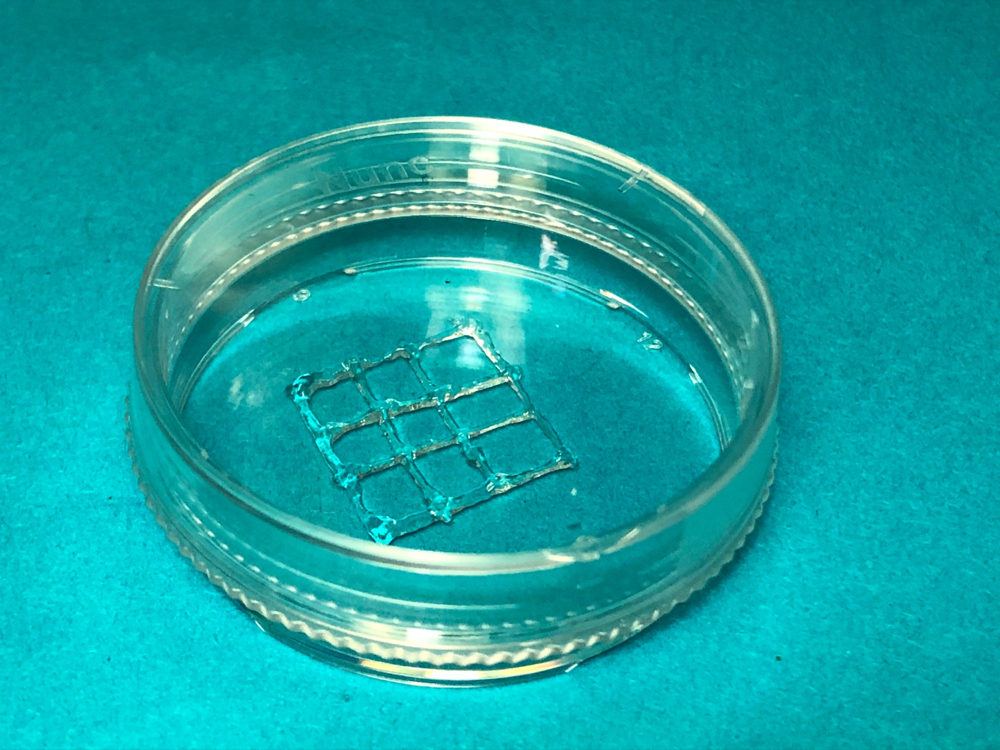
Bioprinted gel on a lab dish. Credit: Wenxiang Zhao
minimally invasive surgical techniques to insert 3-D printing tools into patients through small incisions and then lay down new tissues. Potential applications for such “in vivo bioprinting” might include surgical meshes to help repair hernias and patches for ovaries to help reverse infertility.
Much of the previous research on in vivo bioprinting has focused on treatments of skin and other tissues in the outer part of the body, because the necessary equipment is normally too large to access the digestive tract and other centrally located organs without extensive surgery. In their effort to treat stomach lesions less invasively, scientists in China wanted to develop a miniature bioprinting robot that could enter the human body with relative ease. The researchers used existing techniques for creating dexterous electronic devices, such as
mechanical bees and cockroach-inspired robots, says the study’s senior author Tao Xu, a bioengineer at Tsinghua University in Beijing.
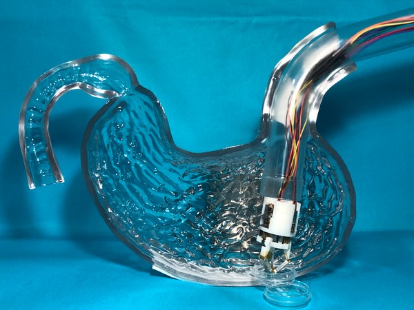
The resulting micro robot is just 30 millimeters wide—less than half the width of a credit card—and can fold to a length of 43 millimeters. Once inside a patient’s body, it unfolds to become 59 millimeters long and can start bioprinting. “The team has constructed clever mechanisms that make the system compact when entering the body yet unfurl to provide a large working area once past the tight constrictions at entry,” says David Hoelzle, a mechanical engineer at the Ohio State University, who did not take part in the study.
In their experiments, the researchers in China fitted the micro robot onto an endoscope (a long tube that can be inserted through bodily openings) and successfully snaked it through a curved pipe into a transparent plastic model of a stomach. There, they used it to print gels loaded with human stomach lining and stomach muscle cells (which were grown in culture by a commercial laboratory) onto a lab dish. The printed cells remained viable and steadily proliferated over the course of 10 days. “This study is the first attempt to combine micro robots and bioprinting together,” Xu says.
The researchers say that mainstream gastric lesion treatments include medications, which can work slowly and are not always very effective; endoscopic surgery, which can only mend relatively small wounds; and endoscopically delivered sprays that staunch bleeding but are of little help in completely healing a larger injury. The hope for in vivo bioprinting is that it might eventually improve on these methods by patching over gastric lesions with living structures that can repair them, Xu says.
Future research could bring the micro robot down to 12 millimeters wide and equip it with cameras and other sensors to help it perform more complex operations, Xu adds. He and lead study author Wenxiang Zhao of Tsinghua University detailed
their findings this summer in Biofabrication.
Xu and his colleagues note that the gels they used as bioprinting “ink” were only stable when relatively cool. At normal body temperatures, they were too liquid to form structures well. Moreover the calcium chloride solution the researchers added to help solidify the gels could potentially damage the human body. But another gel, recently developed independently by Hoelzle and his colleagues, may help address these problems:
it can hold its shape at body temperature and can be solidified using visible light.
One challenge with bioprinting is how toeffectively attach printed cells to existing soft organs and tissues. Hoelzle and his colleagues tested a potential solution by trying to “heal” punctures in texturally similar materials—including raw chicken breast strips. First, the 3-D printer’s nozzle extruded a tiny knob of bio-ink into the puncture, creating an anchor that could connect the pierced tissue to a bioprinted structure. Then they slowly withdrew the nozzle, trailing behind a strand of material they could use to lay more cells on the outside of the tissue. “This work is enlightening,” Xu says. Drawing on these methods, he adds, will help the further development of in vivo bioprinting.
Hoelzle suggests that the technology will likely never become capable of printing complex organs. Instead it may prove useful by augmenting standard surgeries with relatively modest printed structures that could release drugs to promote healing or prevent infection. “There are many opportunities for tissue engineering materials … that are currently not considered—because no one would want to open up the patient to deliver the material,” Hoelzle says.