Back in 2012 some of Ed Boyden’s synthetic neurobiology graduate students at Massachusetts Institute of Technology made a joke: Instead of struggling with slow, bulky and expensive imaging technologies to magnify the tiny neurons in their mouse brain samples, why not make the brains themselves bigger?
Such musings proved prescient when Boyden and his students later stumbled on a polymer that swells (similar to a substance found in the cores of baby diapers), and is able to absorb 200 to 300 times its mass in water. Over the next few years Boyden’s group tinkered with this material to create a new technique called expansion microscopy (ExM), and used it to successfully expand mouse brain samples to about four times their normal size. This allowed them to see structures down to 70 nanometers—roughly the diameter of a flu virus—with common, light-based microscopes (whose lower limit is normally about 200 nanometers).
Now the group has deployed ExM to help another field plagued by a reliance on complex machines to see tiny things: pathology, or the diagnosis of clinical disease using human tissue and blood samples. “We pathologists love to look at things with our own eye,” says renal pathology specialist Astrid Weins, seated in her tiny, windowless office at Boston’s Brigham and Women’s Hospital with a traditional optical microscope on her desk. “We like the old-fashioned way. It’s a trust thing: You don’t trust anything if you don’t see it with your own eyes.”
On supporting science journalism
If you're enjoying this article, consider supporting our award-winning journalism by subscribing. By purchasing a subscription you are helping to ensure the future of impactful stories about the discoveries and ideas shaping our world today.
As the scale of our understanding of the human body has progressed down to a size that eludes human sight, however, scientists like Weins have found themselves forced to rely on increasingly complicated microscopy systems to study cells, molecules and even atoms for signs of disease. That ability comes at a price, literally: An electron microscope (which bounces a beam of electrons, instead of visible light, off the object being viewed) is capable of visualizing structures on the nanoscale—up to 2,000 times smaller than a microscopic bacterium—but can cost as much as $500,000, making it prohibitively expensive for many hospitals and clinics. A top-of-the-line light-based microscope costs around $20,000.
Boyden and his team reported recently in Nature Biotechnology that they are using ExM to enlarge samples that aid disease diagnosis, a new technique they call “expansion pathology” (ExPath). Weins has already used ExPath to see and analyze a special kind of kidney cell—called a podocyte—for specific physical changes that indicate the presence of minimal change disease (an illness that causes protein to be lost through urine) with an optical microscope. She had previously been able to identify those alterations only with an electron microscope. “ExPath is cool because you do see these tiny things with your own eyes—you can see molecular signatures and changes that we couldn’t see before,” Weins says. That ability may open up the routine use of nanoscale imaging in pathology, enabling improved diagnosis and investigation into the basis of disease, she adds.
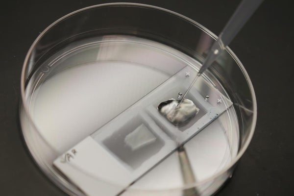
The technology expands samples by first infusing them with a soup of molecules, then chemically triggering the molecules to glom together in a polymer gel that clings to biomolecules within the samples. Next an enzyme is added to loosen up the sample, breaking the bonds that hold the biomolecules together to allow the sample to expand up to four times its original size when water is added. Not only are expanded samples easier to see because they are larger and more transparent, fluorescent tags and other labels can also be added to track individual molecules of interest, including specific genes in the nuclei of cells—a feature electron microscopy lacks. Expansion does not change the spatial arrangement of the biomolecules in a sample as it swells, making it easier to determine where they are in relation to one another. All of that costs a fraction of electron microscope imaging and other “super-resolution” techniques that allow images to be taken at a resolution higher than what is attainable using visible light. The higher resolution is achieved either by tweaking how the light is allowed to bounce off the sample or by using some other source of illumination such as electron beams, laser beams, x-rays or ultrasound waves.
In order to prove their expansion technique works on human pathology samples, the research team’s biggest hurdle was working with the weird ways that researchers prepare tissues for diagnosis. Some are frozen, for example, whereas others are embedded in paraffin. By tweaking their ExM protocol, the team successfully expanded samples from eight different organs (breast, prostate, lung, colon, pancreas, kidney, liver and ovary) by about four and a half times, enabling nanoscale structures such as filaments of keratin and vimentin—proteins that have been associated with metastatic cancer—to be identified with a regular light microscope. When seven observers (including Weins) were asked to analyze images of expanded, fluorescently labeled kidney podocytes, they correctly classified the samples as healthy or indicative of minimal change disease with 90 percent accuracy, compared with only 66 percent accuracy with unexpanded images.
Such an accomplishment is “technically very cool” but unlikely to be widely adopted in clinics immediately, says David Rimm, a Yale University School of Medicine pathologist who was not involved in the study. “Super-resolution microscopy in general is expensive and time-consuming, and we don’t use it much for treating patients. Minimal change disease represents [way less than] 1 percent of anatomic pathology diagnoses done today. So while ExPath is great for that condition, it’s not really clinically relevant yet. However, I look forward to reading many papers in the future that use this technology because I think it’s a better research tool than electron microscopy.”
Rimm might not have long to wait. Boyden and his co-authors have also demonstrated that ExPath can improve a computer artificial intelligence algorithm’s ability to distinguish nuclei in healthy versus cancerous breast tissue. It might take some time to convince the conservative field of pathology to trust computers in addition to human sight, but the future of diagnosis could involve human pathologists and computers working together to analyze tissue samples—their respective performances both enhanced by extending ExM to pathology. “There’s all sorts of unpredictable impacts [of ExM] that are emerging, because there's a huge pent-up demand for a way of looking at three-dimensional things with nanoscale precision,” Boyden says. “This paper has demonstrated that the kind of information we can get from it is quite powerful, so it’s essentially telling the medical imaging community, ‘Look, there could be treasure here. Go hunt.’”