Babies and children undergo massive brain restructuring as they mature, and for good reason—they have a whole world of information to absorb during their sprint toward adulthood. This mental renovation doesn’t stop there, however. Adult brains continue to produce new cells and restructure themselves throughout life, and a new study in mice reveals more about the details of this process and the important role environmental experience plays.
Through a series of experiments, researchers at the Leloir Institute in Buenos Aires showed that when adult mice are exposed to stimulating environments, their brains are able to more quickly integrate new brain cells into existing neural networks through a process that involves new and old cells connecting to one another via special helper cells called interneurons.
The adult mammalian brain, long believed to lack the capacity to make new cells, has two main areas that continuously produce new neurons throughout life. One of these areas, the hippocampus (which is involved in memory, navigation, mood regulation and stress response) produces new neurons in a specialized region called the dentate gyrus. Many previous studies have focused on how the dentate gyrus produces new neurons and what happens to these neurons as they mature, but Alejandro Schinder and his colleagues at Leloir wanted to go one step further and understand how new neurons produced by the dentate gyrus are incorporated into the existing neural networks of the brain, and whether environment affects this process.
On supporting science journalism
If you're enjoying this article, consider supporting our award-winning journalism by subscribing. By purchasing a subscription you are helping to ensure the future of impactful stories about the discoveries and ideas shaping our world today.
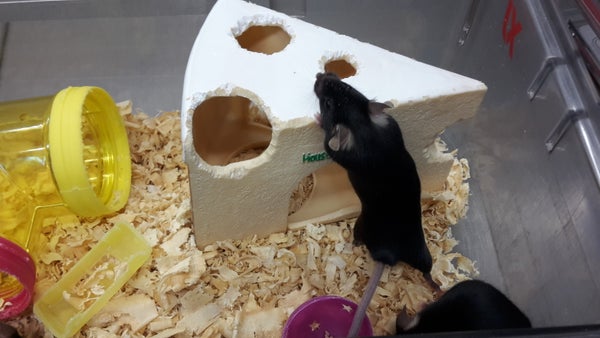
The researchers placed adult mice in an enriched environment, a cage filled with enticing, novel objects such as clear crawl tubes, chew toys and a shelter carved to look like a block of Swiss cheese. As the mice explored, their dentate gyri activated. “They have to create a map of the new environment,” Schinder explains, “So this is a spatial learning process.”
Next, the researchers injected the mice with a harmless virus that labeled the new neurons produced by the dentate gyrus with a red fluorescent protein. Over the next two weeks they placed groups of mice in an enriched environment for different two-day periods; after three weeks they compared the new neurons of the enriched mice to those of control mice that lived in an unadorned cage. The researchers found the new neurons in the enriched mice looked structurally different—they had longer dendrites (the neurons’ input cables that collect information from other parts of the brain) and more spines (locations where neurons connect to one another), which indicate faster growth. Additionally, an analysis of the electrical properties of these new neurons revealed they were more connected to their surrounding cells. According to Schinder, these experiments show “exposing the mice to the enriched environment for two days is sufficient to trigger a process in the cell that will push the cell to connect faster to the preexisting network.”
Armed with this knowledge, the researchers set out to understand the mechanics of how new neurons connect. First they gave some of the mice a treatment that made existing neurons hyperactive for a two-day period, and found that this caused the new neurons to connect to the existing network more quickly, just as when mice experienced an enriched environment. Then they established that intermediate cells, called parvalbumin interneurons, are responsible for conveying signals from old neurons to the new ones, to connect the latter to the existing network. The researchers found that when they made the interneurons hyperactive for two days without placing mice in an enriched environment, the new neurons still connected to the existing network more quickly. When they silenced the interneurons completely but placed the mice in an enriched environment, the new neurons did not connect any more quickly than they did in the control mice. “This is important because now we are saying that the experience that the animal is undergoing is translated to the new cells through these parvalbumin interneurons,” Schinder explains. The results suggest “the process of neurogenesis, which is putting whole new neurons into the hippocampus, is sensitive to really subtle stimuli such as going from a very boring environment to a very rich environment.” The complex environment makes the neurons connect faster, and may prime them for encoding new information.
Hongjun Song, a neuroscientist at Johns Hopkins University who was not involved in the study, finds the results very interesting. His own research has shown these same interneurons play a role in controlling the activation of stem cells and regulating new neuron survival. He says the big takeaway is that everyday experiences can continuously shape physical brain structure. “I think that’s striking,” he says. He adds that the work may have implications for brain disorders such as epilepsy or Alzheimer’s. “What's intriguing is that this type of neuron is vulnerable to disease,” he says, “So I think this paper not only shows how normally an experience can impact the circuit formation continuously in the brain, but also hints at how brain disorders in cells can also cause abnormal regulation of the process.”
Heather Cameron, who studies neuroplasticity at the National Institute of Mental Health and also was not part of the research, is impressed by the way the study “takes things people have said in different contexts—for instance, that interneurons are the first to connect to the young neurons or that enriched environment affects the development or affects neurogenesis, and puts them all together in one complete picture of how this circuit affects new neuron development.”
And although the study is in mice, not humans, Cameron points out that there are a lot of brain similarities between the two species. “We know that the anatomy of the hippocampus and its inputs and outputs are very similar in the two species, and we know that there is ongoing neurogenesis in humans,” she says, “So it's a good bet that these mechanisms seen in mice are also occurring in humans.”