When the now-famous neurological patient Henry Molaison had his brain’s hippocampus surgically sectioned to treat seizures in 1953, science’s understanding of memory inadvertently received perhaps its biggest boost ever. Molaison lost the ability to form new memories of events, and his recollection of anything that had happened during the preceding year was severely impaired. Other types of memory such as learning physical skills were unaffected, suggesting the hippocampus specifically handles the recall of events—known as “episodic” memories.
Further research on other patients with hippocampal damage confirmed recent memories are more impaired than distant ones. It appears the hippocampus provides temporary storage for new information whereas other areas may handle long-term memory. Events that we are later able to remember appear to be channeled for more permanent storage in the cortex (the outer layers of the brain responsible for higher functions such as planning and problem-solving). In the cortex these memories form gradually, becoming integrated with related information to build lasting knowledge about ourselves and the world.
Episodic memories that are intended for long-term storage accumulate to form the “autobiographical” memory that is so essential for our sense of identity. Neuroscientists know a lot about how short-term memories are formed in the brain but the processes underlying long-term storage are still not well understood.
On supporting science journalism
If you're enjoying this article, consider supporting our award-winning journalism by subscribing. By purchasing a subscription you are helping to ensure the future of impactful stories about the discoveries and ideas shaping our world today.
A new study published this month in Science, from neuroscientist Susumu Tonegawa and a group of colleagues at the RIKEN–MIT Center for Neural Circuit Genetics, provides insight into what happens in the brain when a long-term memory is formed, highlighting the critical role of the forward part of the cortex. “It’s the most detailed circuit analysis of the contribution of the prefrontal cortex to memory retrieval we have to date,” says neuroscientist Stephen Maren of Texas A&M University in College Station, who was not involved in the work.
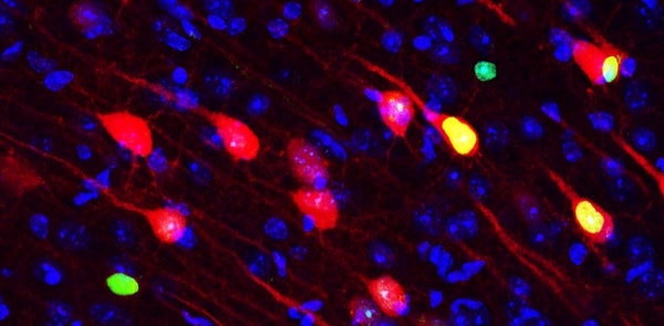
The new study from Tonegawa's group builds on previous research demonstrating that episodic memories are physically represented in populations of cells in parts of the hippocampus. In those studies, the researchers genetically engineered mice so that certain neurons produced light-sensitive proteins. Electrical and chemical activities in the neurons could then be activated or switched off by pulses of light delivered via a fiber-optic cable implanted in each mouse’s skull, a technique known as optogenetics.
The wired mice were given a drug that blocks production of the light-sensitive proteins. Taking the mice off the drug allowed cells that fire while they explored a new environment to make the proteins, effectively “tagging” the memory for that environment. These groups of cells, known as memory “engrams,” could then be controlled with the fiber-optic beams.
With these tools in hand, the investigators gave mice electric shocks to their feet in some of their enclosures, but not others. The mice froze when put back into an environment in which they were previously shocked, indicating a “fear memory.” When the researchers activated the engrams, this invoked the same fearful reaction. The emotional aspects of memories are stored separately, in a region called the amygdala—but activating the engram in the hippocampus activates all linked components, bringing back the full memory. This is similar to how a sound or smell can trigger expansive recall of a past experience in one's life.
In the new study the researchers trained mice to associate a specific cage with foot shocks. Then their memory of what happened was tested on different days up to three weeks later. The researchers tagged engram cells in the cortex and then activated them with light, causing the mice to freeze in environments in which they had never been shocked. The team found these cortical engrams could not be activated by natural cues (being placed back in the enclosure where they were shocked) two days after training, but they could be activated by natural cues 13 days afterward.
This finding shows that although cortical engrams form immediately, they are initially in what Tonegawa calls a “silent” state, meaning they cannot be activated by natural cues. The engrams only mature two weeks later into an “active” state, in which they can respond to such cues. In contrast, hippocampal engram cells were activated by natural cues on the second day after receiving a foot shock, but not on day 13—indicating engrams in the hippocampus become active immediately, but gradually fade into a “silent” state.
Tonegawa’s research points to the existence of complementary memory systems: One allows rapid memory formation but has limited capacity, and thus needs to pass information that should be retained to another system that is longer-lasting but slower-acting. This frees space in the hippocampus that can then be reused. “There is a division of labor. The hippocampus can form active memories very quickly, while the cortex takes care of long-term stability,” Tonegawa explains. “If you don’t need prolonged memory, the hippocampus is enough; if you don’t have to form active memory quickly, the cortex is enough; but we want both.”
The findings help clarify when and how cortical memories are formed. One prior theory held that information is slowly transferred into the cortex, but Tonegawa’s findings support the alternative idea that cortical engrams are formed immediately but need time to develop. “The key question this work resolves is whether memory engrams move from the hippocampus to cortical storage sites over time or are established in the cortex during learning and unmasked as time passes,” Maren says. “This is strong evidence for the latter.”
The team also showed that blocking inputs to the amygdala from the hippocampus during memory testing impaired short-term memory performance (tested on the second and eighth days)—but not distant memory (tested on days 15 and 22)—whereas blocking inputs to the amygdala from the cortex showed the opposite pattern. In other words, memory engrams in the amygdala were maintained throughout, and were necessary for recalling fear memories—but there was a change in which region the amygdala needed to be connected to in order for memory to function. “The cells that allow a mouse to remember the fear [aspect] of a memory are maintained from day 1 to three weeks later,” Tonegawa says. “But there's a switch in the use of connections: at three weeks, when the hippocampus engram is not active anymore, the connectivity between the prefrontal cortex engram and the amygdala engram allows the animal to recall the fear memory.”
The study “provides compelling evidence about where and when specific neurons contribute to a particular form of memory in an essential way at particular times during and after learning,” says neuroscientist Howard Eichenbaum, director of the Center for Memory and Brain at Boston University, who was not part of the research. Even though it is a technical tour de force, the paper leaves several open questions: “The study doesn’t tell us whether there are other cells that are important for this kind of memory, or anything about other types of memory,” he says. Most important, he adds, it only tells us that certain cells, in some regions at specific times, make memories—not how they contribute to doing so. “What kind of information processing does the prefrontal cortex contribute,” he says, “that is not essential for learning or retrieval shortly after but which becomes essential some time later?” Each region involved has different functions and processes information in different ways. None are specifically dedicated to memory, which consists of traces left behind as these neural systems process experiences. The hippocampus, for instance, represents spatial information using “place” cells that map the environment, potentially explaining how it might contribute to the “where” component of episodic memory. It is not clear what role prefrontal cortex processing plays but Eichenbaum speculates its role in organizing and choosing between alternatives might be increasingly relevant as memories age.
A more incisive picture of the way memory works is slowly starting to emerge and these new findings will help propel further research into various kinds of memory.