If you stumbled across a radio or a computer and had no idea how it worked, you would likely first want to find out what it was made from—what its component parts were. Your next step might be to determine what each component actually did, taking care to note which parts were connected to others.
This is the approach to understanding the brain taken in two related studies published October 31 in Nature. The first used differences in the activity of genes to identify different cell types in mouse brains, finding more distinct types than previously known. The second took a close look at a few of the identified cell types to try to work out what they do, showing they perform two quite different roles in controlling movement.
The first study, led by molecular geneticist Bosiljka Tasic of the Allen Institute for Brain Science, set out to catalogue the different brain cell types in two parts of the mouse cortex (the brain's outer layer, responsible for higher cognitive functions). The researchers chose two distant regions: the primary visual cortex, the first stop for processing visual input from the eye, and part of the motor cortex (anterior lateral motor cortex, or ALM), which is involved in movement. They analyzed 23,822 cells using a technique that allowed them to determine which of over 45 thousand possible genes were actually active in individual cells. They then grouped cells with similar genetic activity together to identify 133 different cell types. “This is the most comprehensive description of cortical composition we have to date,” Tasic says.
On supporting science journalism
If you're enjoying this article, consider supporting our award-winning journalism by subscribing. By purchasing a subscription you are helping to ensure the future of impactful stories about the discoveries and ideas shaping our world today.
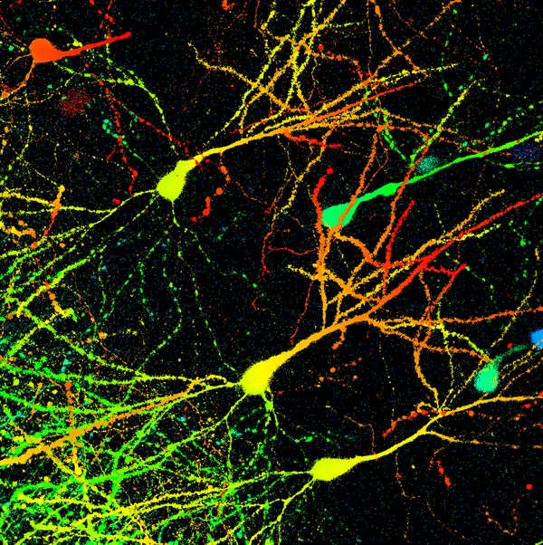
The cell types fall into two broad classes: One, called inhibitory neurons, communicates using the chemical messenger GABA and inhibits the activity of other, nearby cells. The other class, excitatory neurons, uses the chemical glutamate and they serve as the brain's main cells for generating neural activity. A major finding of the study was that different types of inhibitory neurons (the team identified 61 inhibitory subtypes) are almost all shared between the two regions studied. In contrast, 51 of the 56 excitatory subtypes identified were unique to the region they were found in. (Sixteen cell types that were not neurons were also found) “This is a landmark study that reveals previously unappreciated [diversity] of excitatory cortical neurons,” says neurobiologist Tomasz Nowakowski of the University of California, San Francisco, who co-authored an accompanying commentary but was not involved in the study. “These were long presumed to be largely similar across cortical areas. Tasic and colleagues demonstrate this is far from truth.”
The team next used a fluorescent marker to trace connections going to distant brain regions from over 2,000 cells. “Inhibitory neurons have local connections that modulate what happens within the area they work in,” Tasic says, “while most excitatory neurons project to distant places, which could be other cortical areas, or noncortical areas.” They found neurons with different patterns of long-range connections also had different gene activity profiles, providing further confirmation the categories they identified are distinct cell types, likely to play different roles in brain function.
The second study, led by neuroscientist Michael Economo of Howard Hughes Medical Institute’s Janelia Research Campus in Virginia, focused in on a few subtypes identified in the first study, to try to understand their functions. Tasic and colleagues found three subtypes in a specific layer of the ALM, which contains excitatory “pyramidal tract” neurons. “We were really interested in these,” Economo says. “Because they’re the main output neurons of the motor cortex, connecting it with important motor centers elsewhere responsible for initiating and carrying out movement.” (The pyramidal tract neurons are also the ones that degenerate in some motor neuron diseases, such as ALS. So understanding the information these cells transmit and the circuitry they are part of could help researchers better understand what goes awry.)
Economo and colleagues found two of the subtypes connect to the thalamus, a central “hub” region that links back to the cortex, forming a loop. The other subtype connects to the medulla, a brain stem region containing neurons that activate muscles and coordinate movement. This arrangement tallies well with previous work suggesting a distinction between neurons involved in communicating preparatory movement— “plans”—and those that initiate and control movement. “A lot of fast, voluntary, flexible movements involve some preparation period before they’re performed,” Economo says. “This allows animals to perform movements faster and with more precision once they're initiated.”
Having established this circuitry, Economo and colleagues conducted experiments in mice to pin down what the neurons actually do. They trained mice to respond to having their whiskers poked in one of two places by drinking from a spout to either their left or right. While the mice performed this task the researchers recorded the activity of cells in the ALM using a long, flexible electrode. The researchers had labeled the neurons they were interested in using optogenetics, (a technique that makes cells photo-sensitive and thus allows them to be easily identified by shining light on them). They could then observe the cells’ activity during a motor task to study their roles in movement. Results showed some cells connected to the medulla were highly active at the start of a movement, whereas others switched on either in the midst or end of the activity.
Cells connected to the thalamus, on the other hand, displayed a complex, sustained pattern of activity as soon as the mice's whiskers were poked, but before they moved. “There's a stable code there for exactly what the stimulus was and what kind of movement the mice are going to perform,” Economo says. “This is what we expect a motor plan to look like—a representation of the upcoming movement that persists over time.”
These findings show three of the cell types identified in the first study play two quite different roles: one involved in preparing movements, the other in initiating and controlling them. “The answer’s so clear and beautiful,” Tasic says. “It's a perfect showcase of how you can use this type of data.” This covers just three of the 133 cell types thus far identified, but the atlas of genetic identities provided by the first study, which is openly available to other researchers, will enable researchers to zero in on other types to conduct similar experiments. “These studies set out a vision for the future of neuroscience: to comprehensively characterize all major cell types in the brain and systematically define their function,” Nowakowski says. “It also shows we’re only just at the beginning of this journey.”