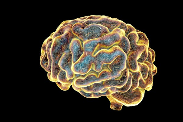
Imagine the following transformation. A pea-sized chunk of your skin breaks apart in a dish of salts and serums. The mixture is infected with viruses that make some cells smaller, more circular, and clump together. They’ve turned into stem cells. Then, a bath of other salts, serums, and factors coax them into becoming mature neurons. The neurons divide and organize themselves into three dimensional spheres. Inside the spheres, the neurons layer themselves like the neurons in your cerebral cortex. There’s not just one ball, but an army of tiny spheres. Each sphere contains thousands of neurons; each neuron with a copy of your DNA. The neurons communicate with each other with pulses of electricity. The spheres start to organize structures that look a lot like the different lobes and substructures of your brain. Some of the spheres may even form an optic cup, an early version of your retina. This may seem like a perverse form of human cloning carried out by a neuroscientist turned witch-doctor. But it’s real: an emerging laboratory model system that might one day help treat you or a loved one’s debilitating neurological disorder.
They are called brain spheroids (or three-dimensional brain cultures or cerebral organoids) and are a relatively new creation. They were first described in a splashy study published in Nature in 2013 and are one of the most technically impressive forms of tissue culture.
What brain spheroids are not, however, is as important as what they are. They’re not ‘mini-brains’. They’re not generating thoughts and emotions. Without any sensory input they lack grounding in the physical world. Brain spheroids are also very small. At 4 mm in diameter, they’re much smaller than a mouse’s brain. They’re this small because real developing brains need massive amounts of nutrients throughout the depth of their structure. Brain spheroids get their nutrients from a bath of serums. But without a network of blood vessels to deliver the serum to deeper parts of the spheroid, these parts starve. Attaching a functioning circulatory system to the spheroids isn’t feasible with current techniques, so making bigger, more developed spheroids seems unlikely at this point.
On supporting science journalism
If you're enjoying this article, consider supporting our award-winning journalism by subscribing. By purchasing a subscription you are helping to ensure the future of impactful stories about the discoveries and ideas shaping our world today.
Still, these 4 mm brain spheroids can be useful as is—or with some custom modifications. A recent paper in Nature Methods illustrates this point by using brain spheroids to study an important cell-type in your brain called oligodendrocytes and a rare disease associated with their dysfunction. Oligodendrocytes form a fatty sheath called myelin that insulate axons, the long protrusions your neurons use to communicate to other neurons. Think rubber insulation around a copper wire. The electrical messages traveling along axons need myelin to both maintain an electrical charge and to transmit the message quickly. By adding a few steps to the standard brain spheroid method, the authors made spheroids that contain both neurons and oligodendrocytes that ensheath the neurons’ axons, just like in the brain.
These oligocortical spheroids, as they’re dubbed, more closely resemble a real developing brain. But more importantly, the authors showed that oligocortical spheroids can be used to study disorders that impair oligodendrocyte development and myelination. First, the authors treated their oligocortical spheroids with two FDA-approved “pro-myelination” drugs and saw accelerated oligodendrocyte development and myelination in the spheroids. Next, they made new spheroids where stem cells harbored genetic mutations seen in Pelizaeus-Merzbacher disease, a rare inherited disorder that causes a deficit in myelin formation during development. The prognosis for Pelizaeus-Merzbacher disease is poor and current treatments are merely palliative. The authors showed that the mutation-carrying spheroids were deficient in myelin formation, mimicking Pelizaeus-Merzbacher pathology. Remarkably, a not-yet-approved drug improved myelination in one of these disease-mimicking spheroids.
This finding is promising news for families supporting a loved one with Pelizaeus-Merzbacher disease. But Pelizaeus-Merzbacher disease is rare—affecting between 1/200,000 and 1/500,000 people. Rare diseases, as a whole, though, are not rare. The Online Mendelian Inheritance in Man (OMIM)—which compiles profiles and statistics on inherited diseases—has an entry for 3,486 inherited neurological disorders. Most of these disorders affect small numbers of patients spread around the globe. The small numbers make coordinating clinical trials nearly impossible. To make matters more complex, rare diseases like Pelizaeus-Merzbacher disease can cause a range of symptoms and severities, with different treatment needs. An alternative would be for healthcare providers to test a drug or combination of drugs on a patient’s individual brain spheroid, made from their very own skin cells.
This technology won’t be in the neurology clinic soon, however. In oncology, which has outpaced neuroscience in developing organoid models, implementation of personalized organoids for clinical practice is still years away. Also, different types of spheroids would need to be tailor-made for different disease processes like oligocortical spheroids were for Pelizaeus-Merzbacher disease. Using a patient’s own cultured tissue to tailor their treatment course is not a new proposal. But, brain spheroids make a new type of personalized medicine feel closer to being achievable in neurology.